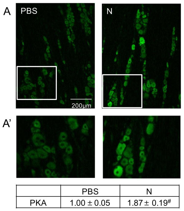
Fig. 5.
Nicotine administration increased expression of PKA in trigeminal ganglion neurons. (A) Representative images of sections from the V1/V2 region of trigeminal ganglia obtained from control (PBS) and nicotine (N)-treated animals immunostained using antibodies directed against PKA. Enlarged images of the ganglion that contained numerous neuronal cell bodies and associated satellite glial cells from panel A are shown (A′). The change in PKA staining intensities are reported as the average fold change±SEM as compared to mean levels in control samples that was set equal to 1 (n=3). #P ≤ 0.01 when compared to control values.

