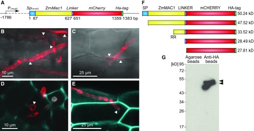
Figure 2.
U. maydis Strain SG200Zmmac1 Secretes Full-Length ZmMAC1 Fusion Protein into the Maize Apoplast.
(A) Schematic overview of the Trojan horse construct integrated into the genome of U. maydis strain SG200 to obtain strain SG200PUmpit2::SpUmpit2-ZmMac1-mCherry-Ha (SG200Zmmac1). The ZmMac1 coding cDNA sequence was fused to mCherry and an HA-tag. To guarantee successful fungal secretion, the coding region was fused at the N terminus with the signal peptide coding region of the U. maydis effector pit2, which targets ZmMAC1 to the maize apoplast (Doehlemann et al., 2011a). Because Umpit2 is highly expressed in planta 2 to 3 dpi, its promoter was used to drive SpUmpit2ZmMac1-mCherry-Ha expression (Doehlemann et al., 2011a).
(B) to (E) Secretion of the ZmMAC1-fusion protein monitored by confocal imaging.
(B) and (C) U. maydis hyphae infecting seedling leaf epidermal cells. SG200Zmmac1 hyphae within epidermal cells of maize seedling leaves. The fusion protein (red) localized around the hyphae and spreads from the fungal hyphae into the apoplast of contiguous cells at sites of cell-to-cell passage (indicated by white arrowheads).
(D) and (E) Transverse section of an anther lobe (D) and anther epidermal cells (E) infected with SG200Zmmac1. The fungal hyphae are growing intracellularly, while the ZmMAC1-fusion protein (red) is secreted and surrounds the fungal hyphae (indicated by white arrowheads). To visualize maize cell walls using autofluorescence (blue), an excitation of 405 nm and detection at 435 to 480 nm were used.
(F) Schematic overview of different ZmMAC1 protein cleavage versions including predicted molecular weight. RR refers to a predicted protease site (Supplemental Figure 2).
(G) Immunodetection by anti-HA serum of ZmMAC1-fusion protein pulled down from SG200Zmmac1 infected spikelet extract with anti-HA or unconjugated agarose matrix after extraction in the presence of protease inhibitors and after formaldehyde incubation of spikelets.

