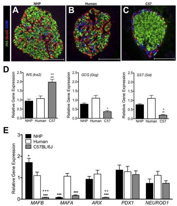
Figure 6. Differences in islet morphology and composition in different species.
Non-human primate islets (A) are more similar in architecture to human islets (B). Both species have less defined mantle and core domains than mouse islets (C). In mice, β cells are restricted to the islet core while other cell types are found at the periphery. In non-human primates and humans, non-β cells are found at the periphery and internal to the islet. (D) β cells comprise a larger proportion of endocrine cells in mouse islets compared with the other two species. (E) Expression of islet transcription factors differs between species. *P < 0.05, **P < 0.01, and ***P < 0.001, mouse vs. human; + P < 0.05 and ++ P < 0.01, +++ P < 0.001 mouse vs. NHP. (Adapted from Conrad, E., Dai, C., Spaeth, J., Guo, M., Cyphert, H. A., Scoville, D., Carroll, J., Yu, W. M., Goodrich, L. V., Harlan, D. M., Grove, K. L., Roberts, C. T. Jr., Powers, A. C., Gu, G. and Stein, R. 2016, Am. J. Physiol. Endocrinol Metab., 310(1), E91–e102 with permission from American Physiological Society.)

