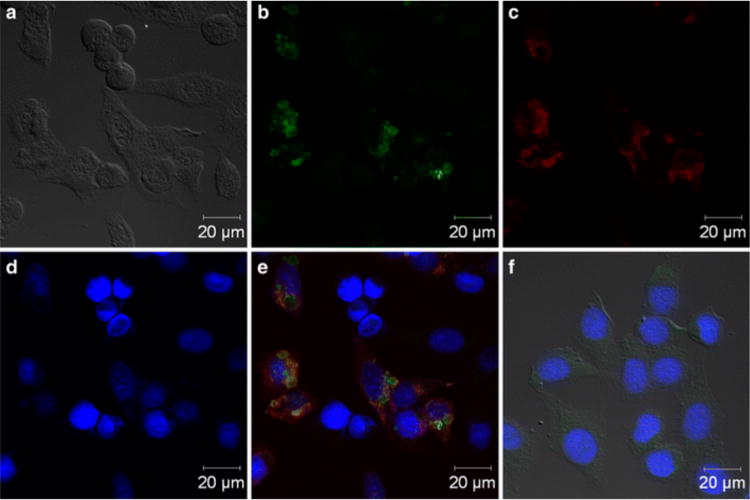
Figure 7.

Peptide TADKLLYGLFKS can be used in the fluorescence labeling of cowpox-infected HEp-2 cells. (a) Bright-field image of cells. (b) Green fluorescent image of cells on glass slides incubated with the peptide and streptavidin–fluorescein isothiocyanate. (c) Cells were also incubated with a polyclonal anti-D8 antibody coupled to DyLight 649 (shown in red) to assess colocalization. (d) The nuclei of cells were stained by DAPI (blue). (e) Overlay of b, c, and d showing all stains. (f) Noninfected stained HEp-2 cells (control) do not show fluorescence.64 Reproduced with permission from ref64. Copyright 2014 Springer.
