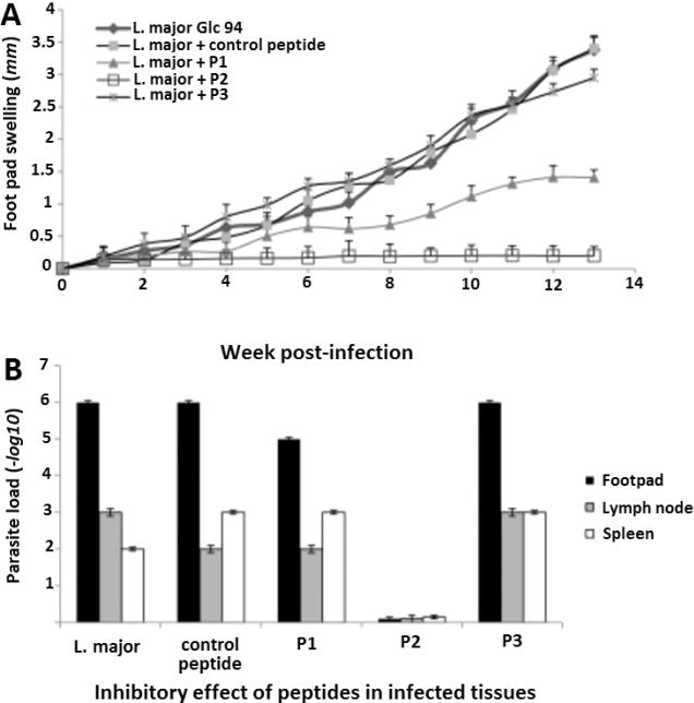
Figure 9.

Inhibitory effect of selected peptides in infected tissues. (A) Inoculation of BALB/c mice with several virus library derived L. major inhibitory peptide candidates. L. major Glc 94 metacyclic promastigotes (1.0 × 10⁶) were preincubated with 100 μM of peptides P1 (MSKPKQ), P2 (MAAKYN), P3 (MAHYSG), or control peptide, or with phosphate-buffered saline (PBS). Next, 5 mice per group were inoculated with these preincubated mixtures. The sizes of lesions were monitored with a Vernier caliper. Lesion sizes were calculated by subtracting the size of the contralateral-uninfected footpad. Swellings were monitored over a 13-week observation period every week. The results shown are the mean lesion size of 5 mice per group in millimeters. (B) Parasite burden in infected footpads. The burden was determined after draining the lymph nodes and spleens of mice by a limiting-dilution assay at week 13 postinfection. Results shown are the means ± the standard deviations of log10 dilutions at the three anatomical sites. These results include triplicates for each group with 5 individuals in a group.51 Adapted with permission from ref51. Copyright 2016 Elsevier.
