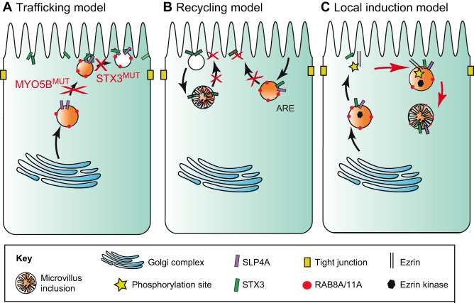
Fig. 3.
Three models to explain the pathological hallmarks of MVID. The panels depict human enterocytes, showing endosomal trafficking routes (black arrows). The apical surface is uppermost. (A) In the trafficking model, defects (depicted by red crosses) in vesicle trafficking (caused by MYO5B mutations, MYO5BMUT) or delivery (caused by STX3 mutations, STX3MUT) result in the subapical accumulation of vesicles and in the lack of appropriately polarized apical proteins. (B) In the recycling model, defects in the recycling and delivery of apical recycling endosomes (AREs) result in the subapical accumulation of apical proteins and in the formation of microvilli-containing macropinosomes. (C) In the local induction model, MVID results in colocalization of ezrin and ezrin kinases in the subapically accumulated AREs to create a signaling platform that results in local ectopic microvillus formation, which leads to the formation of microvillus inclusions (red arrows). In healthy cells, ezrin kinases are transported to the apical membrane where they activate ezrin by phosphorylation to induce microvillus formation.

