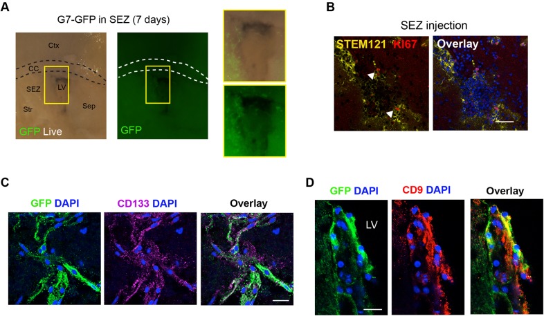
Fig. 2.
Human patient-derived GSCs engraft in adult mouse SEZ. (A) Direct microinjection of human line into adult SEZ and visualization of engrafted live cells using a constitutive GFP reporter. The boxed areas are shown at higher magnification on the right. (B) Immunostaining for human-specific cytoplasmic antigen (STEM121; yellow) and KI67 (red) after 14 days. Arrowheads indicate cells immunopositive for STEM121 and KI67. (C,D), Marker analysis after 21 days of ex vivo culture: immunocytochemistry for CD133 (purple) (C) and CD9 (red) (D). Nuclear counterstaining was performed with DAPI (blue). Scale bars: 50 µm in B; 20 µm in C and D.

