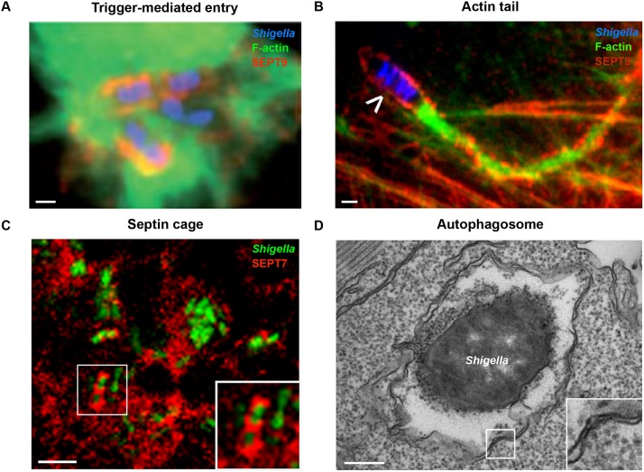
Fig. 2.
The hallmarks of Shigella infection. (A) Trigger-mediated entry by Shigella. HeLa cells were infected with Shigella (blue), fixed for fluorescent microscopy, and labelled with antibodies to SEPT9 (red) and phalloidin for F-actin (green) to highlight septin recruitment at the site of Shigella entry. Scale bar: 1 µm. (B) The Shigella actin tail. HeLa cells were infected with Shigella (blue; white arrowhead indicates a motile bacterium) for 3 h, fixed for fluorescent microscopy, and labelled with antibodies to SEPT2 (red) and phalloidin for F-actin (green) to highlight septin ring formation around the actin tails. Scale bar: 1 µm. (C) The Shigella-septin cage in vivo. SEPT7 (red) assembles into cage-like structures around S. flexneri (green). Zebrafish larvae were infected with green fluorescent protein (GFP)-Shigella for 4 h, fixed, labelled with antibodies to SEPT7 and imaged by confocal microscopy. The inset shows a higher magnification view of the boxed region in C, showing Shigella entrapped within a septin cage. Scale bar: 5 µm. (D) An autophagosome sequestering cytosolic Shigella in vivo. Zebrafish larvae were infected in the tail muscle with GFP-Shigella for 4 h and fixed for electron microscopy. The inset shows a higher magnification view of the boxed region in D, showing the double membrane, a hallmark of autophagosomes. Scale bar: 0.25 µm. Images adapted from Mostowy and Cossart (2009) (A), Mostowy et al. (2010) (B) and Mostowy et al. (2013) (C,D).

