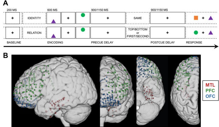
Fig 1. Working memory task design and electrode coverage.
(A) Single-trial working memory task design. Following a 1-s pretrial fixation interval (−250 to −50 ms pretrial epoch used as baseline), subjects were directed to focus on either “identity” or “relation” information. Then, 2 common shapes were presented for 200 ms each in a specific spatiotemporal configuration (i.e., top/bottom spatial and first/second temporal positions). After a 900- or 1,150-ms jittered precue fixation delay, the test cue appeared (i.e., one word presented on screen for 800 ms), followed by a postcue fixation delay of the same length. Working memory was tested in a two-alternative forced choice test (0.5 chance rate). In the identity test (top), subjects indicated whether the pair was the “same” pair they just studied (correct response in this example: “no”). In the spatiotemporal relation test (bottom), subjects indicated which shape fit the top/bottom spatial or first/second temporal relation cue (correct response for cue “top” or “second”: “circle”). (B) Reconstruction of electrode coverage for all subjects. Electrodes are overlaid on the left hemisphere, displayed in 4 views. Red = MTL; green = PFC; blue = OFC. Underlying data can be found in University of California, Berkeley, Collaborative Research in Computational Neuroscience database (http://dx.doi.org/10.6080/K0VX0DQD). MTL, medial temporal lobe; OFC, orbitofrontal cortex; PFC, prefrontal cortex.

