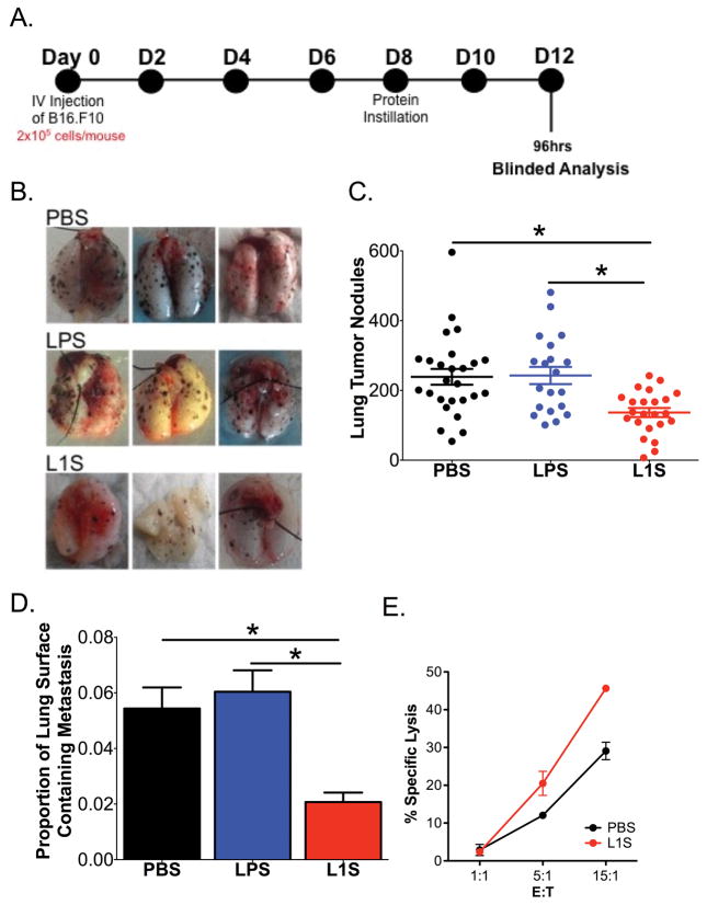
Figure 4. L1S instillation stimulates a therapeutic anti-tumor response in mice with established “metastatic” B16.F10 melanomas.
Mice were given tumor cells on d0 and treated on d8 by instillation with 125 μg of PBS ± L1S or LPS. Mice were sacrificed on d12 (96 h after treatment). (A) Experimental timeline. (B) Representative images of mouse lungs at time of harvest. (C) Plot depicts quantified tumor nodules counted on lungs of the treated mice at harvest. Date are pooled from seven independent experiments. (D) The proportion of lung surface area covered with tumor nodules was quantified as a measure of total tumor size. Data are from the same experiments as in (C). (E) Lysis of RFP-expressing B16.F10 tumor cell targets by NK cells purified from lungs of mice at 24 h after instillation of PBS ± L1S protein. Data are from one of three experiments with similar results. All graphs depict means ± SEM. (*) indicates p < 0.05 using ANOVA with Newman-Keuls comparison.

