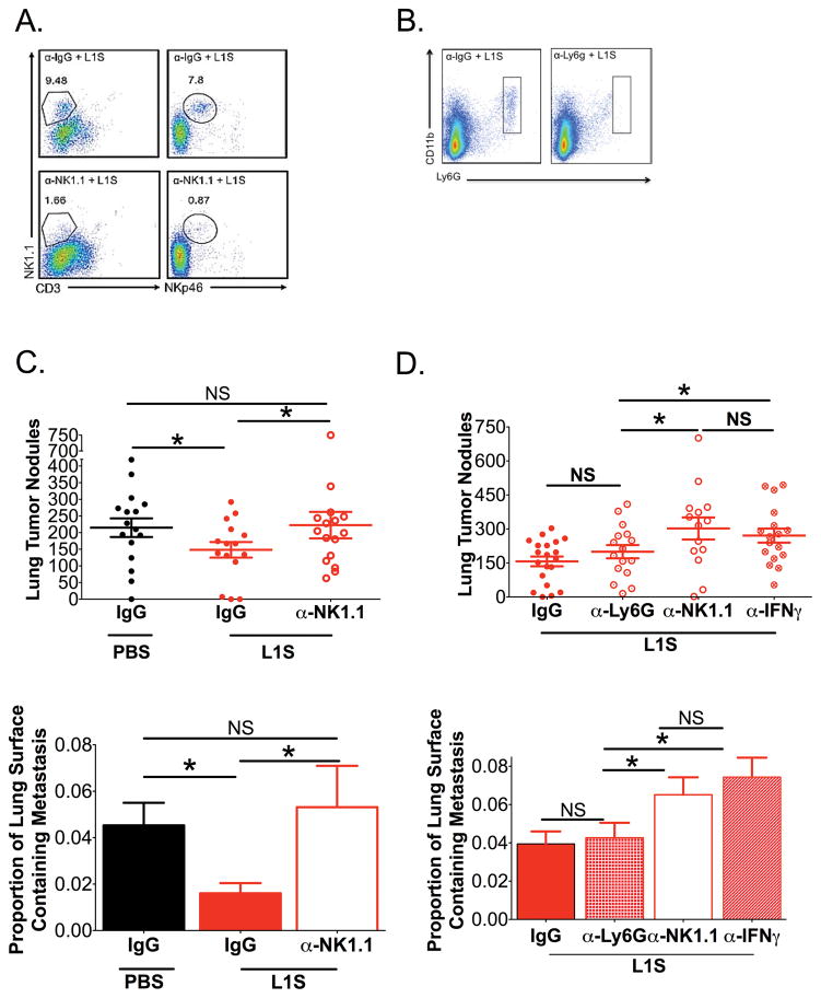
Figure 5. Therapeutic effects of L1S treatment are dependent on NK cells and IFNγ.
Mice were inoculated with B16.F10 cells as above. Groups of these mice received i.p. inoculation with depleting antibodies on d7 and i.t. treatment with PBS± L1S on d8 as indicated. Harvest were performed on d12. (A) Dot plots depict NK cells (NK1.1+CD3−) from lung homogenates of mice treated with a control IgG (a-IgG) or α-NK1.1 by analysis of both NK1.1+CD3− and NKp46+CD3− after instillation of L1S purified by Ni-column chromatography treated mice. (B) Representative FACs dot plots of Neutrophils by analysis of Ly6G+CD11b+ in L1S purified by Ni-column chromatography treated mice given α-IgG control or α-Ly6G depletion. (C) Tumor nodules and tumor area are quantified on the lungs of mice with established tumors and are depleted of NK cells and treated with L1S purified by Ni-column chromatography or PBS (N=4). (D) Tumor nodules and tumor area are quantified on the lungs of mice with established tumors and are depleted of NK cells, IFNγ, or Neutrophils and treated with L1S purified by Ni-column chromatography (N=5). Cells are gated on Live → singlets → Respective group(s) of interest. For all graphs, * indicates p<.05 using ANOVA with Newman-Keuls comparison. Shown are mean ± SEM

