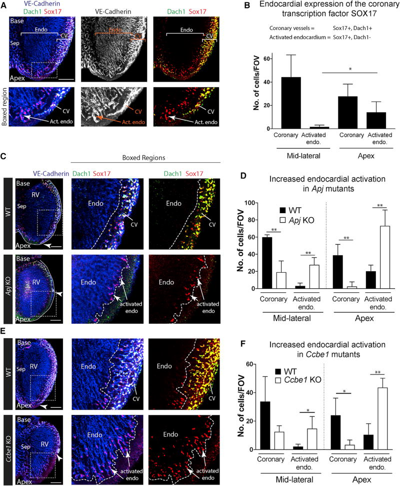
Figure 5. SOX17 Is a Marker of Activated Endocardium that Is Expanded in Mutants with Deficient SV Sprouting.
(A) Immunostaining to label VE-cadherin, DACH1, and SOX17 in E13.5 hearts. DACH1 and SOX17 are widely expressed in coronary vessels (CV); SOX17 is also expressed in a subset of endocardial cells near the compact myocardium at the apex where they produce coronary vessels (activated endocardium).
(B) Quantification of the number of coronary (DACH1+, SOX17+) and activated endocardial (DACH1−, SOX17+) cells in the indicated regions of the heart (n = 6 hearts). Error bars are mean ± SD.
(C–F) SOX17 immunofluorescence in Apj (C and D) and Ccbe1 mutants (E and F) show that activated endocardium is expanded where SV-derived coronary vessels are absent. Arrowheads indicate the leading front of coronary migration. Quantifications shown in (D) and (F) where error bars represent SDs. Apj (n = 4 wild-type, n = 5 KO hearts); Ccbe1 (n = 3 wild-type, n = 4 KO hearts).
Endo, endocardium; Sep, septum; Act. Endo, activated endocardium; RV, right ventricle; CV, coronary vessels; FOV, field of view; WT, wild-type.
*p ≤ 0.05, **p ≤ 0.01. Scale bars, 200 µm. See also Figures S5 and S6.

