FIGURE 3.

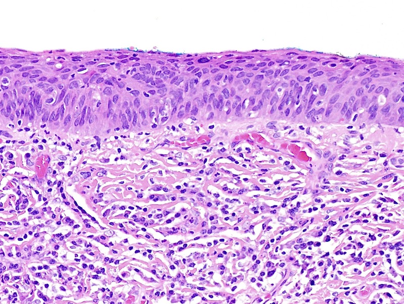
Basaloid dVIN seen as thinned epithelium, full-thickness atypia, stromal fibrosis, and a dense lymphoplasmacytic infiltrate; p16 was negative (H&E ×200).

Basaloid dVIN seen as thinned epithelium, full-thickness atypia, stromal fibrosis, and a dense lymphoplasmacytic infiltrate; p16 was negative (H&E ×200).