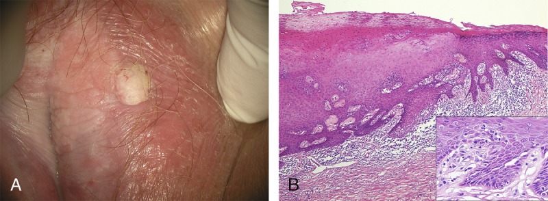
FIGURE 4.

A, A 74-year-old with verrucous SCC, dVIN, and VAAD on background of pallor, lichenification, and architectural change characteristic of LS. B, VAAD adjacent to dVIN is demonstrated by thickened epithelium, parakeratosis, and an underlying pale band, with a sharp transition to the spiky rete ridges and basilar atypia (inset) of standard dVIN. Throughout there is a dense lymphocytic infiltrate and stromal sclerosis (H&E ×40, inset H&E ×400).
