
Fig. 2.
Primary cilia deficiency promotes intracranial aneurysm development in knockout mice.
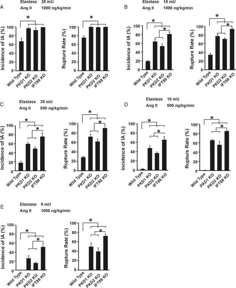
A, Incidence of IA (left panel) and rupture rate (right panel) after 20 days of IA induction with 1,000 ng/kg/min angiotensin-II and 35 mU elastase. n = 12 for each type of mice. ∗ indicates P < 0.05. B, Incidence of IA (left panel) and rupture rate (right panel) after 20 days of IA induction with 1,000 ng/kg/min angiotensin-II and 10 mU elastase. n = 12 for each type of mice. ∗ indicates P < 0.05. C, Incidence of IA (left panel) and rupture rate (right panel) after 20 days of IA induction with 500 ng/kg/min angiotensin-II and 35 mU elastase. n = 12 for each type of mice. ∗ indicates P < 0.05. D, Incidence of IA (left panel) and rupture rate (right panel) after 20 days of IA induction with 500 ng/kg/min angiotensin-II and 10 mU elastase. n = 12 for each type of mice. ∗ indicates P < 0.05. E, Incidence of IA (left panel) and rupture rate (right panel) after 20 days of IA induction with 1,000 ng/kg/min angiotensin-II. n = 12 for each type of mice. ∗P < 0.05. PKD1 indicates polycystin 1; PKD2, polycystin 2; IFT88, intraflagellar transport 88; KO, knockout; Ang II, Angiotensin II; IA, intracranial aneurysm.