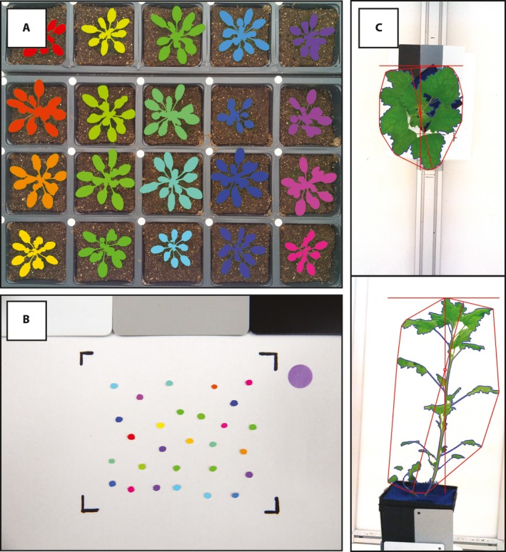
Figure 2.

Examples of data collected from Raspberry Pi phenotyping platforms that have plant and/or seed tissue segmented using open‐source open‐development software PlantCV (Fahlgren et al., 2015). (A) PlantCV‐segmented image of a flat of Arabidopsis acquired from Raspberry Pi time‐lapse imaging protocol in a growth chamber. (B) PlantCV‐segmented image of quinoa seeds acquired from Raspberry Pi camera stand. (C) Example side‐ and top‐view images of quinoa plants acquired from Raspberry Pi multi‐image octagon. Plant convex hull, width, and length have been identified with PlantCV and are denoted in red.
