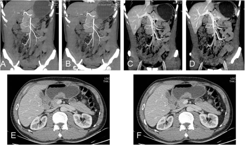
Figure 3.

CTE of a 53-year-old male patient (BMI: 20.1 kg/m2) with gastric adenocarcinoma underwent CTE at 80 kVp/270 mg I/mL with FBP and 50%ASIR algorithm. Maximum intensity projection images of SMA reconstructed with FBP (A), 50% ASIR (B) and SMV reconstructed with FBP (C), 50% ASIR (D). Axial images show wall thickening of the gastric antrum reconstructed with FBP (E) and 50% ASIR (F) respectively. CTDIvol, DLP, ED, and total iodine dose were 7.18 mGy, 316.67 mGy-cm, 4.75 mSv, and 16.85 g. SD of the images reconstructed with 50% ASIR was reduced obviously and subjective image quality was similar between the 2 reconstruction methods.
