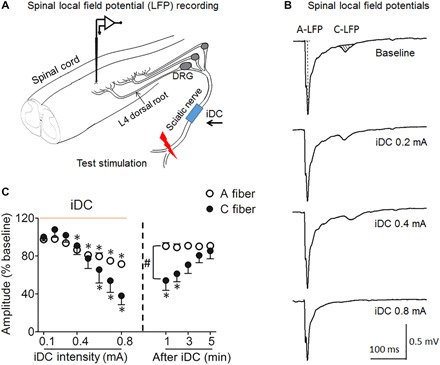
Fig. 4. Cathodic iDC at the sciatic nerve suppresses spinal LFP to peripheral test stimulation in an intensity-dependent manner.

(A) Experimental setup for recording LFP from the superficial dorsal horn at the L4 spinal level to a test pulse (25 V, 0.5 ms) applied at the distal sciatic nerve in rats. Monopolar cathodic iDC stimulation was applied to the sciatic nerve at mid-thigh level. (B) Example traces show spinal LFP evoked by test stimulation before and after iDC stimulation. LFPs corresponding to A fiber and C fiber activation were distinguished on the basis of latency. The peak amplitude of A-LFP and area under the curve (AUC; shaded area) of C-LFP were measured off-line. (C) The amplitude of A-LFP and AUC of C-LFP decreased progressively as amplitudes of iDC increased (0.1 to 0.8 mA, 2 min per amplitude) and gradually recovered during the first 5 min after iDC cessation.*P < 0.05 versus pre-iDC baseline; #P < 0.05 versus the indicated group at post-iDC, two-way repeated-measures ANOVA with Tukey post hoc test. Data are mean + SEM (n = 8).
