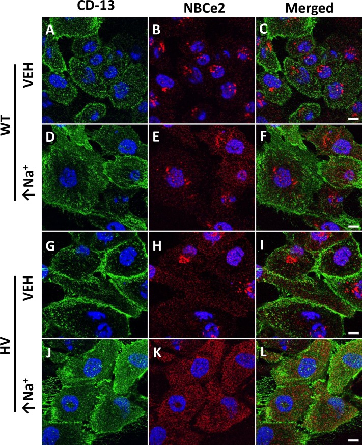
Fig 2. Immunofluorescence localization of NBCe2 in human renal proximal tubule cells (hRPTCs) carrying wild-type (WT) or homozygous variant (HV) SLC4A5 imaged on Petri dishes using confocal microscopy.
CD-13, a specific membrane-bound ectopeptidase present in RPT but not in other nephron segments, in the membrane is stained green using a more photostable fluor (CD-13-Alexa 488 antibody) (A, D, G, and J), NBCe2 is stained red (B, E, H, and K), nucleus is stained blue in all the images, including merged images (C, F, I, and L). With monensin treatment (10 μmol/L 24 hr) which increases intracellular sodium (↑Na+), NBCe2 expression gets more diffuse similar to the apical membrane stain (E and F, K and L), especially for HV cells. Panels C, F, I, and L show merged images of NBCe2 and CD-13; there is increased NBCe2 on the surface in HV cells treated with monensin (K and L). The scale bar in panel C, F, I, and L = 10 μm.

