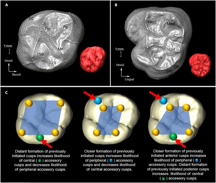
Fig. 1. Anatomical and developmental configuration of molar cusps.

Occlusal view of the EDJ of an (A) upper and (B) lower molar with cusp nomenclature. (A) Paranthropus robustus (SK831a, ULM3) and (B) Australopithecus africanus (STW560b, LLM3). An occlusal view of outer enamel surface (OES) is shown in red as an inset (not to scale). pro, protocone; pa, paracone; me, metacone; hyp, hypocone; C5, cusp 5; CC, Carabelli’s cusp; prod, protoconid; med, metaconid; end, entoconid; hyd, hypoconid; hyld, hypoconulid/cusp 5; C6, cusp 6 (tuberculum sextum); C6D, double cusp 6; C7, cusp 7. The OES in (A) exhibits both cusps 5 and 6 (cusp 6 not present at EDJ). (C) Schematic of potential developmental pathways during tooth crown morphogenesis. Location of EKs (yellow spheres) superimposed onto an OES and exaggerated for visualization purposes. Note that in developing teeth, cusp spacing is regulated by EKs, affecting the growth of the intercuspal regions and distances (denoted by blue polygons) at which new EKs and cusps form. Growth of tooth after the cusp patterning can further modify the cusp distances.
