
Figure 3. Illustration of local tissue inhomogeneity around nerve leading to transients in drivers of activation.
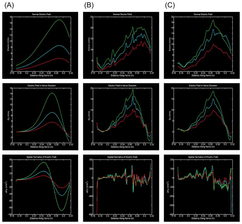
(A) 0.180 m (B) 0.217 m (C) 0.250 m (D) 0.263 m (E) 0.269 m. Five anatomical cross sections showing cases in which either fat and soft tissue (A, B, C, E) or just fat (D) borders the vagus nerve. Slices (B, C, D, E) are relatively close to the stimulating electrodes while slice (A) is relatively far.