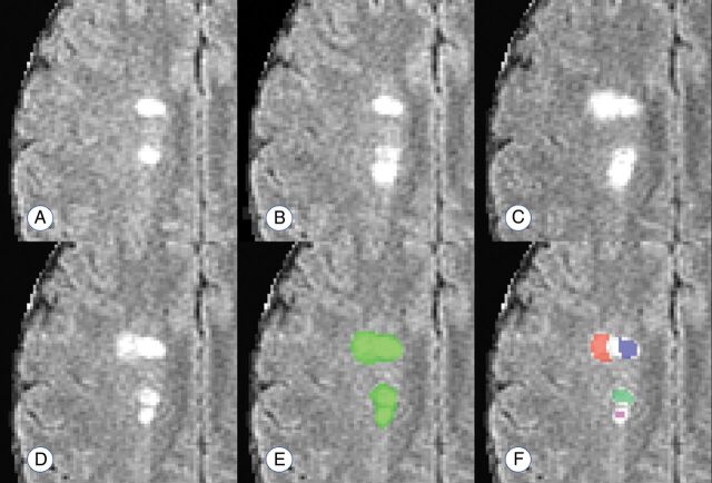
Fig 2.
Example of the lesion counts in a region with 4 apparently distinct lesions, 2 of which develop with observable temporal separation. A–D, Development of 2 new and temporally distinct lesions. E and F, The performance of a connected-components count and the proposed count, respectively. The connected-components method finds 1 confluent lesion in the visualized space (connected in an adjacent plane), and the proposed method finds 4 distinct lesion centers. Days from scan in A: 28 days (B); 91 days (C); 252 days (D–F).

