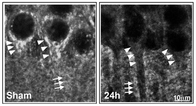
Fig. 2.
Confocal microscopic images of CA1 neurons labeled with NSF antibody. Brain sections were obtained from a sham-operated control rat and rat subjected to 15 min of ischemia followed by 24 h of reperfusion. NSF is located in the peri-nuclear and dendritic truck (Sham, arrowheads), as well as in the neuropil (Sham, arrows) of sham-operated control CA1 neurons. NSF is mostly depleted from the CA1 neuronal perinuclear region and dendritic trunk (24h, arrowheads), but remains relatively at the sham control level in the CA1 neuropil (24h, arrows) at 24 h of reperfusion after transient cerebral ischemia.

