
Fig.4. Effects of 0.2 μm LPS with and without RS-LPS 2.0 μm on β-APP axon transport analyzed by Western blotting assays.
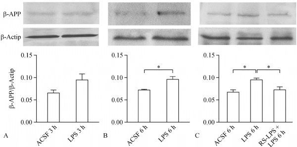
A: β-APP density was obviously enhanced by 3 hours incubation of LPS, but this did not reach to a significant difference (P= 0.089, t-test) vs. the control. B: β-APP accumulation was significantly higher in CC tissues vs. the control when duration of LPS treatment was doubled to 6 hours (P< 0.05, *,t-test). C: treatment of the CC slices with LPS induced significant accumulation of β-APP compared to the control (P< 0.05, *, by one-way ANOVAwith Sidak's posttest); meanwhile, co-incubation of LPS and RS-LPS for 6 h significantly reversed β-APP density increment compared to LPS treatment alone (P< 0.05, *, with Sidak's posttest).