
Fig.2. Microscopic pictures of fungal growth.
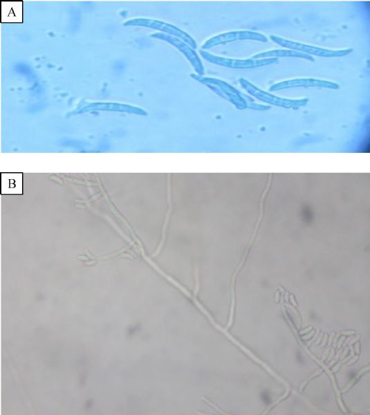
A: LPCB mount showing septate hyphae and sickle shaped macroconidia (3-8 ×11-17 μm) with 3–4 septae under 40× magnification. B: Septate branched hyphae with lateral monophialide conidiophore under 40× power.