
Figure 3.
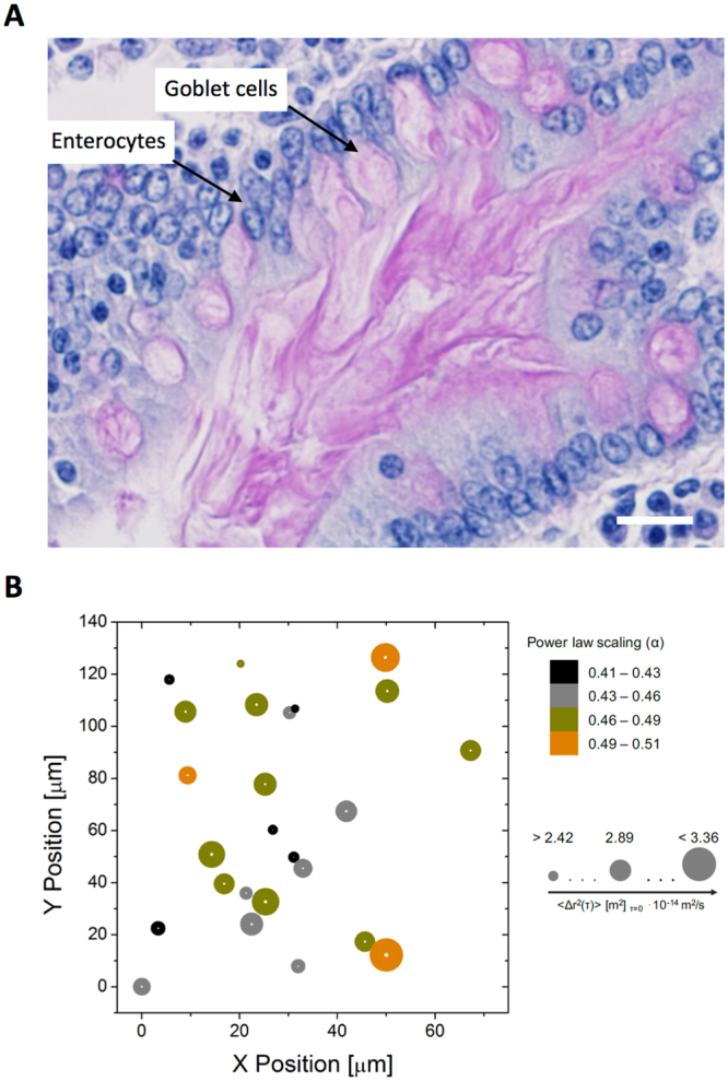
A structural model of mucus comprising rheologically heterogeneous domains blended together. (A) Periodic acid-Schiff staining of the pseudostratified columnar epithelium from the proximal small intestinal porcine mucosa which illustrates the presence of domains in physiological mucus. Note the pink staining of mucus from goblet cells into the intestinal lumen and the blue staining of nuclei. Scale bar 20 μm. (B) Visualization of the rheological heterogeneity, X- and Y-positions, of movement of individual 0.5 μm colloidal particles in 50 mg/mL ND-mucin solution. The size of circles indicates diffusivity and the color corresponds to the power law scaling of the MSD spectra.