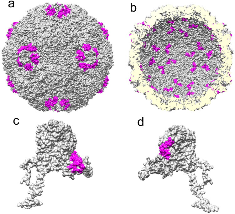
Figure 2.
RCAP of capsid protein VP1. RCAP results of recombinantly-expressed VP1 are shown on the HPeV1 model (PDB: 4Z92). (a) Peptides from VP1 identified by RCAP that are localized to the outer capsid surface (b) Peptides from VP1 identified by RCAP that localize to the inner capsid surface. (c) Locations of the peptides that contact RNA on a monomer of VP1 with a view corresponding to the outer capsid surface whereas (d) A view of VP1 that corresponds to the inner capsid surface.

