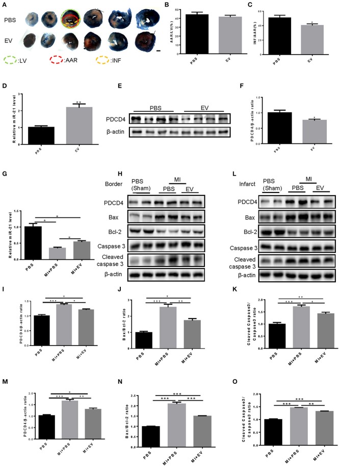
Figure 6.
Mouse serum extracellular vesicles increased miR-21 levels, reduced the AMI-induced infarct area and attenuated cardiomyocytes apoptosis in mouse. (A) Photographs of TTC staining of six slices from mice cardiac tissues (n = 4). Scale bar, 1,000 μm. (B,C) Analysis of AAR/LV and INF/AAR ratio to define the infarct area of hearts from mice treated with MI and EVs extracted from mice serum (n = 4). (D) qRT-PCRs for expression of miR-21 in mouse heart tissues at 24 h after the injection of EVs. (E,F) Western Blot for expression of PDCD4 in mouse heart tissues at 24 h after the injection of EVs. n = 4; (G) Quantitative real time-polymerase chain reactions (qRT-PCRs) for expression of miR-21 (n = 4). (H–K) Western blot of border tissues for PDCD4, Bax, Bcl-2, Caspase 3, and Cleaved caspase 3, and quantitative analysis for PDCD4, Bax/Bcl-2 and Cleaved caspase 3/Caspase 3 ratio. β-actin was used as a loading control (n = 4). (L–O) Western blot of infarct tissues for PDCD4, Bax, Bcl-2, Caspase 3 and Cleaved caspase 3, and quantitative analysis for PDCD4, Bax/Bcl-2, and Cleaved caspase 3/Caspase 3 ratio. β-actin was used as a loading control (n = 4). *0.01 < P < 0.05; **0.001 < P < 0.01; ***P < 0.001. EVs, Extracellular vesicles.

