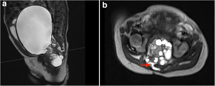
Fig. 1.
Preoperative, T2-weighted sagittal (left), and axial (right) MR images of the abdomen and pelvis, demonstrating a large cystic mass within the pelvis abutting the sacrum and coccyx. A small posterior extension of the pelvic mass, extending through an enlarged sacral foramen, is observed posterior to the sacral elements (arrow)

