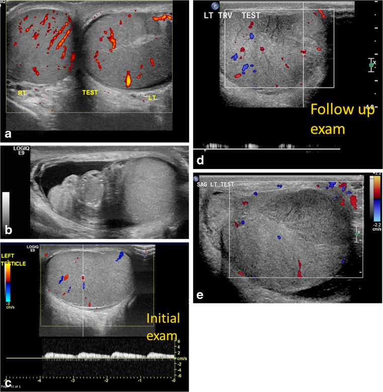
Fig. 9.
Partial torsion in a 13-year-old boy who presented with a 2-week history of intermittent left testicular pain and swelling. a Transverse power Doppler US image of both testes shows bilateral preserved flow with a slightly globular-appearing left testis. b Gray-scale longitudinal US image of the left testis and epididymis shows an enlarged left epididymal-cord complex and surrounding small hydrocele. The boy was discharged with a diagnosis of epididymitis because of the long confounding duration of symptoms and presence of intratesticular flow. c Color and pulsed Doppler US image of the left testis at initial presentation shows preserved intrinsic flow with normal arterial waveform. d Follow-up exam after 3 weeks demonstrates abnormally dampened left testicular flow and waveforms. e Color Doppler US image of left testis during at follow-up for persistent pain shows interval significantly enlarged left testis with decreased vascularity and a focal hypovascular parenchymal area concerning for infarct in the setting of incomplete torsion. He underwent left detorsion with bilateral orchiopexy

