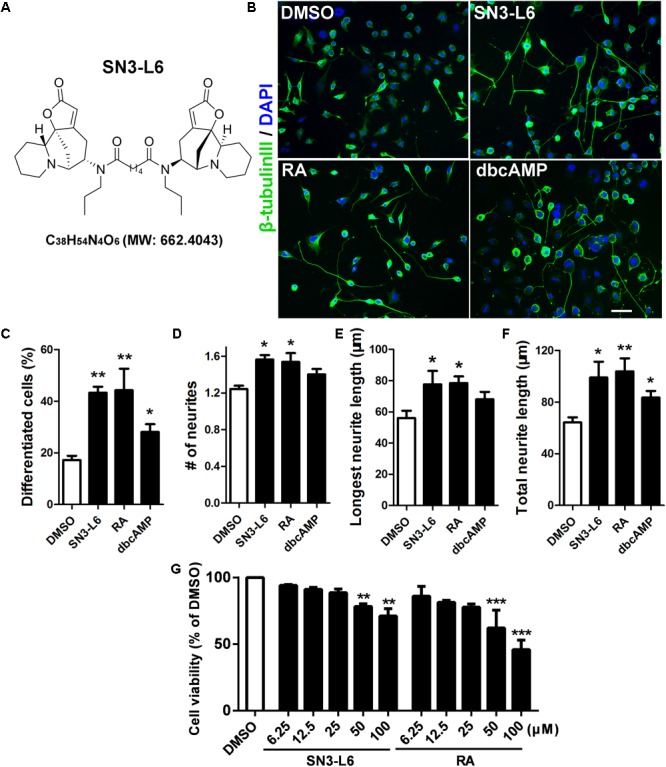
FIGURE 1.

SN3-L6 potently promotes differentiation and neurite outgrowth. (A) The chemical structure, the chemical formula and the exact mass of SN3-L6 are shown. (B) Representative images of Neuro-2a cells after treatment with SN3-L6 (25 μM), RA (10 μM), or dibutyl cyclic AMP (dbcAMP) (0.5 mM) for 48 h. Scale bar, 50 μm. Neurites and nuclei were visualized using β-tubulin III antibody (green) and 49,6-diamidino-2-phenylindole (DAPI) (blue), respectively. Differentiation rate (% of cells that possess at least one process longer than 40 μm; C), neurite number (D), longest neurite length (E), and total neurite length (F) were quantified. ∗P < 0.05, ∗∗P < 0.01, indicated compound vs. DMSO. (G) Neuro-2a cells were treated with SN3-L6 or RA in a concentration gradient (6.25, 12.5, 25, 50, and 100 μM) for 48 h. Cell viability was tested using MTT assay. ∗∗P < 0.01, ∗∗∗P < 0.001, SN3-L6 or retinoic acid (RA) vs. DMSO. All data shown in this figure are presented as mean ± SEM from at least three independent experiments. Statistical analysis was subjected to one-way ANOVA with Bonferroni multiple comparison test.
