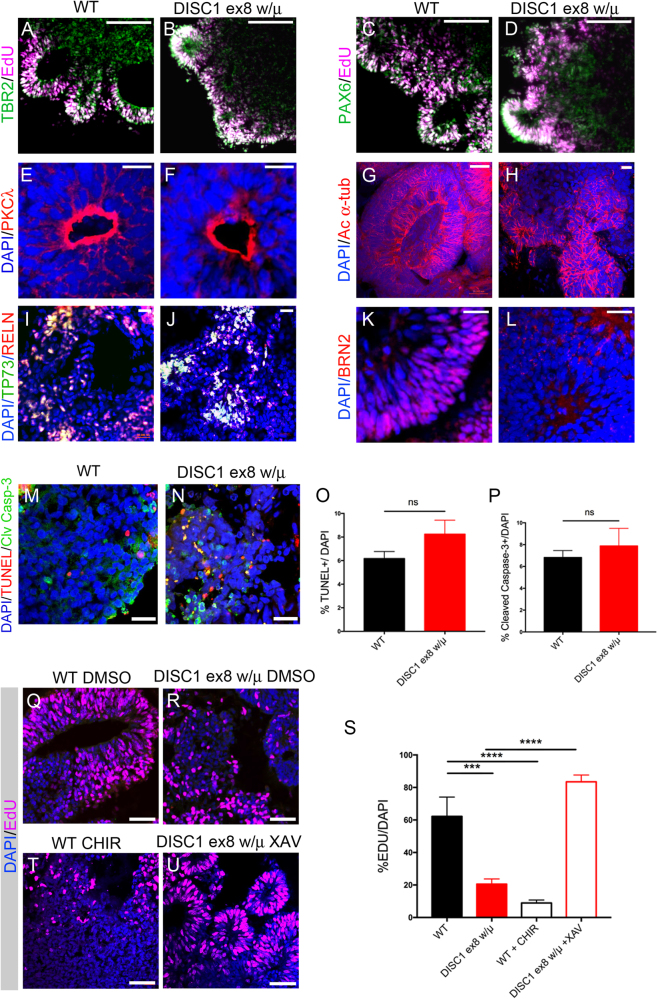
Fig. 3. DISC1-mutant organoids exhibit decreased BRN2 expression and reduced proliferation that is rescued by WNT antagonism.
Immunostaining was performed on WT and DISC1-mutant day 19 organoids for EdU incorporation and markers as shown. Expression of cell fate markers TBR2, PAX6, P73, Reelin and polarity markers PKC-λ, and acetylated-α-tubulin were grossly unchanged (a–j). However, immunostaining of BRN2 was markedly reduced in DISC1-mutant organoids (k, l). Scale bars: a–d 100 μm, e–h 20 μm, i–j 50 μm, k–l 20 μm. m, n WT and DISC1 exon 8 wt/μ organoids were immunostained at day 19 for markers of apoptosis (TUNEL and Cleaved Caspase 3). o, p Quantification of percentage of DAPI positive nuclei positive for TUNEL or cleaved caspase-3 shows no difference with DISC1 disruption. q–u WT and DISC1-mutant organoids with WNT agonism (CHIR) or WNT antagonism (XAV) were pulsed with EdU for 2 days (culture days 6–7), then fixed, sectioned, and stained for EdU and DAPI at day 19. One representative image for each condition is shown. s Percentage of EdU-positive nuclei were quantified. Data were derived from three independent differentiations. Statistics: o, p Variance was significantly different between conditions, Welch’s t-test. s Variance was significantly different between conditions, one-way ANOVA with Geiser–Greenhouse correction, Sidak’s multiple comparisons test. ***p < 0.001, ****p < 0.0001. Scale bars: m, n, q, p, t, u: 50 μm

