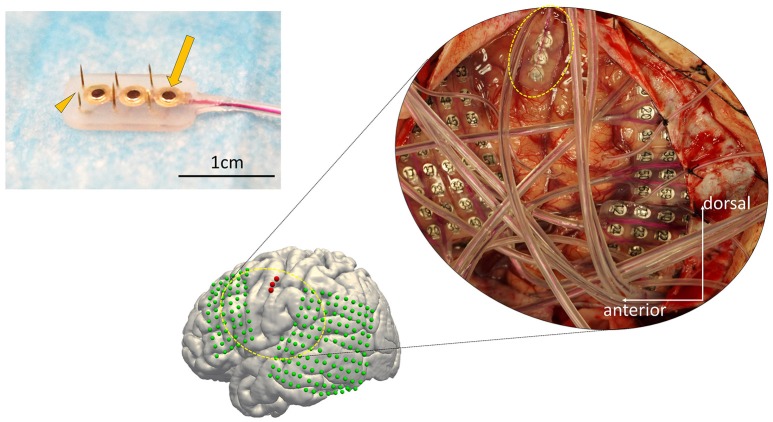
Figure 1.
The configuration of the hybrid electrode and an example of implantation. (Left) The electrode fabricated for this study (7 × 13 mm) consists of three grid electrodes (macroelectrodes, arrow) and six needle electrodes (microelectrodes, arrowhead) on the side (Unique Medical Co., Tokyo, Japan). The surface diameter of each macroelectrode was 1.5 mm, whereas that of the microelectrodes was 200 μm. The length of the needles alternated between 1.5 and 2.5 mm to reduce the physical force needed for implantation. (Right) Implanted electrodes overlaid on a 3D reconstructed pre-operative MRI, displayed together with the intraoperative photograph. The hybrid electrode is encircled by the dotted yellow line in the intraoperative photograph. The subdural grid electrodes are shown in green. The hybrid electrode is shown in red.

