
FIGURE 1.
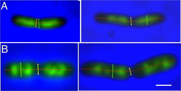
Examples of deeply-constricted Escherichia coli MC1000 cells, fixed with osmium tetroxide and stained with DAPI. Fluorescence images are overlaid with phase-contrast images and with markers, transiently displayed upon the images, for cell length (red), cell diameter (green) and the diameter at the constriction site (yellow), as measured by “Coli-Inspector” with ImageJ plugin ObjectJ. (A) Slow growing cells in succinate medium (Td = 122 min). Left and right panel, cell shorter and longer than 3.7 μm, respectively. (B) Fast growing cells in glucose plus amino acids medium (Td = 29 min). Left and right panel, cell shorter and longer than 4.3 μm, respectively. See mean lengths in Table 1, column 10. DAPI fluorescence is seen in green. Scale bar equals 1 μm.