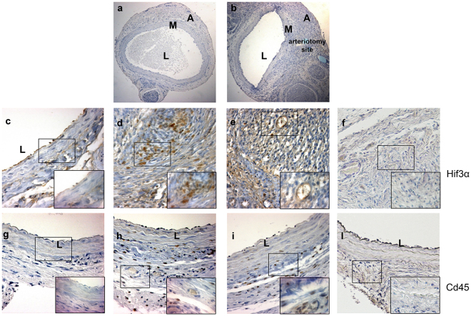
Figure 5.
Immunohistochemical analysis of Hif3α and Cd45 expression in uninjured rat carotids and in carotids from Wistar male rats treated with MSCs or DMEM and harvested 7 days after arteriotomy. (a) Uninjured rat carotid; (b) arteriotomy-injured rat carotid harvested 7 days after injury, haematoxylin staining. Arrows in b indicate the injury site, where arteriotomy is followed by the application of an 8.0 polypropylene stitch (light blue). (c–f) Representative immunohistochemical staining of Hif3α in uninjured rat carotid (c) and in injured carotids harvested 7 days after arteriotomy from DMEM- (d) and MSC-treated rats (e). (g–l) Representative immunohistochemical staining of Cd45 in adjacent cross-sections from uninjured rat carotid (g) and in injured carotids harvested 7 days after arteriotomy from DMEM- (h) and MSC-treated rats (i). (f,l) Immunohistochemical staining of serial cross-sections of rat carotids used in d and h without primary antibody as negative control. (a,b) 10x magnification; (c–l) 20x magnification; small insets: 40x magnification of selected areas enclosed in black rectangles, representative of nuclei positive to Hif3α and Cd45 in adventitia, vasa vasorum and perivascular tissue. Brown staining corresponds to target protein expression. Nuclei were counterstained with haematoxylin. L: lumen; M: media; A: adventitia.

