Fig. 3.
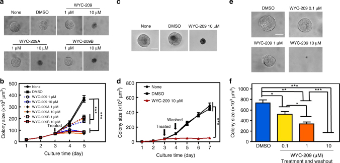
Inhibition of tumor cell colony growth by WYC-209 with or without washout. a Murine melanoma cells were cultured in 90-Pa fibrin gels for 3 days and then treated with medium alone (None), DMSO (0.1% DMSO-containing medium), 1 or 10 μM WYC-209, 1 or 10 μM WYC-209A, WYC-209B. Representative images of colonies on day 5; Scale bar, 50 μm. b Summarized data. Mean ± s.e.m.; n = 15 samples; three separate experiments; ***P < 0.001. c Representative images of colonies on day 7: None: untreated; DMSO: treated with 0.1% DMSO as a vehicle; WYC-209 10 μM: 3 days after WYC-209 was washed out; Scale bar, 50 μm. d Drugs were added on day 3 and washed out on day 4; the cells were cultured till day 7. Mean ± s.e.m.; n = 15 samples; three separate experiments; ***P < 0.001. e WYC-209 abrogates colony formation of TRCs. DMSO: Control B16-F1 cells, cultured in 90-Pa 3D fibrin gels and treated with 0.1% DMSO on day 0 for 5 days, were then re-plated into 90-Pa 3D fibrin gels as single individual cells for 5 days without treatment. WYC-209 0.1 μM, 1 μM, or 10 μM: Control B16-F1 cells, cultured in 90-Pa 3D fibrin gels and pretreated with WYC-209 on day 0 with various concentrations for 5 days, then after washing out WYC-209, the cells were re-plated into 90-Pa 3D fibrin gels for 5 days. Representative images of colonies on day 5 (all colonies started as a single cell on day 0); Scale bar, 50 μm. f Summarized colony sizes on day 5. Mean ± s.e.m.; n = 15 samples; three separate experiments; *P < 0.05; **P < 0.01; ***P < 0.001. Student’s t-test was used in all statistics

