Figure 3.
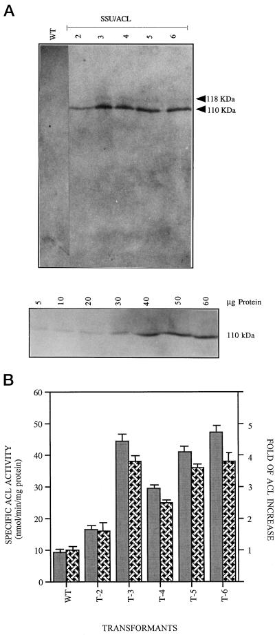
Expression of ACL in tobacco leaves. A, Protein extracts, 50 μg, of crude plastid fractions were loaded on 6% (w/v) SDS-PAGE, blotted on membranes, and probed with rat ACL antibody. Lane 1, Untransformed cells (wild type, WT); lanes 2 to 6, SSU/ACL transformants T2 to T6, respectively. The 110-kD band corresponds to the mature ACL protein. Bottom panel, Plastids isolated from an SSU/ACL transformant (T6) were treated with thermolysin and different amounts of proteins (5–60 μg) were loaded in each lane and probed with anti-ACL antibody. An increased intensity of signal was observed with an increase in the amount of proteins electrophoresed in individual lanes. B, Comparison of ACL activity (gray bars) and relative increase of ACL activity (hatched bars) among the SSU/ACL transformants T2 to T6 compared with the activity in the original, untransformed wild type (WT) cells. Bar represents the mean of three separate assays.

