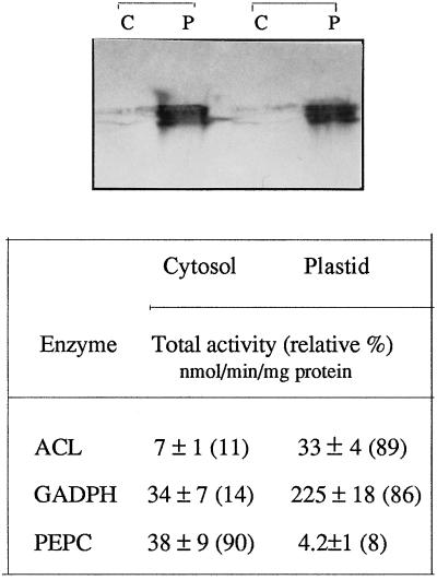
Figure 4.
Localization of ACL protein in plastids. About 30 μg of protein extracts of both plastid (P) and cytosol (C) fractions were separated on SDS-PAGE and the resulting gel was immunoblotted with rat ACL antibody. The majority of ACL proteins localized in the plastidic fraction can be observed. This experiment was repeated with identical results (duplicate is shown in top panel). Bottom panel, Purity of plastid and cytosol fractions based on the activities of NADPH-glyceraldehyde-3-P dehydrogenase (as a plastidic marker) and PEP carboxylase (as a cytosolic marker). The relative percentage of activity is shown in parentheses.

