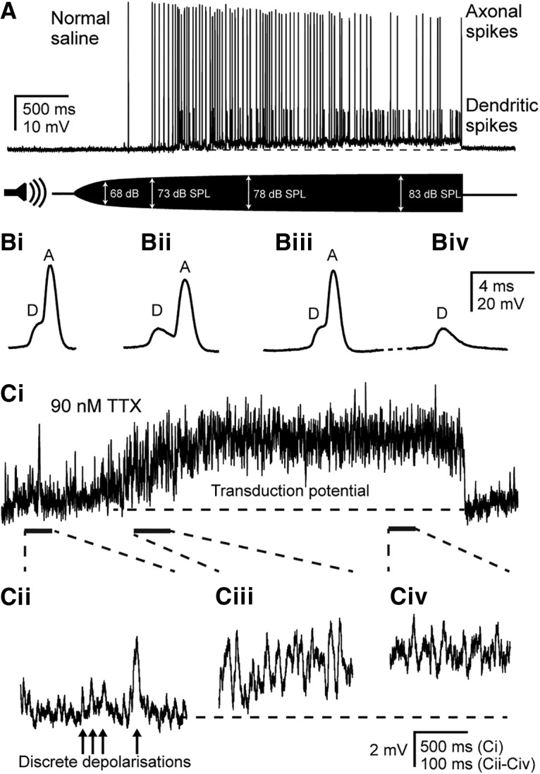
Figure 3.

Dendritic spikes trigger axonal spikes, and discrete depolarizations summate to produce a graded transduction potential. A, Current-clamp recording from the soma of a Group III neuron at a resting membrane potential of − 75 mV. The cell is depolarized and produces both small-amplitude dendritic spikes and large-amplitude axonal spikes in response to a 3 kHz tone that is amplitude ramped from 0 to 83 dB SPL. Bi–Biv, Expanded view of spikes in A at progressive time points during the recording. Dendritic spikes precede and give rise to axonal spikes (Bi–Biii), but axonal spikes can fail, leaving only the underlying dendritic spike (Biv). Ci, Transduction potential in response to the same amplitude-ramped 3 kHz tone as shown in A, but with 90 nm TTX to block spikes, measured at resting potential (note different vertical scale compared with A). Cii–Civ, Expanded views of transduction potential at the time points indicated by dashed lines and solid bars, showing discrete depolarizations and their summation to produce a graded transduction potential. Horizontal dashed lines in A and Ci indicate the resting membrane potential of −75 and −69 mV, respectively.
