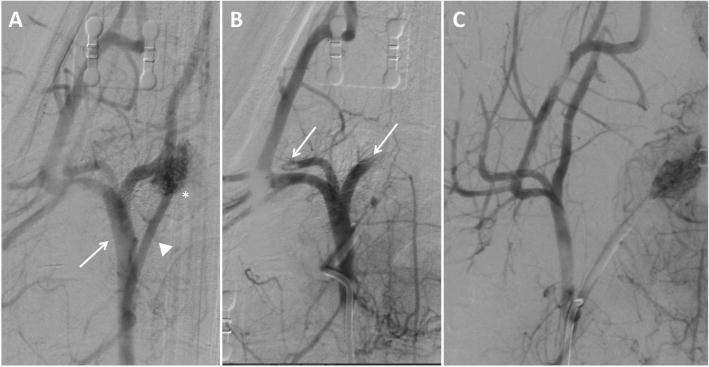
Fig. 6.

Digital subtraction angiography (DSA) imaging of right carotid vasculature from a single experiment. (a) Baseline start of study with the arrow pointing to the external carotid artery (ECA) and the asterisk identifying the ascending pharyngeal artery (APA) supplying the rete mirabile. (b) Post clot occlusion of carotid vasculature showing absent blood flow to the APA and rete mirabile with arrow identifying occlusion of blood flow to branches of the ECA. (c) Post embolectomy through manual aspiration identifying patent ECA, APA, and rete mirabile
