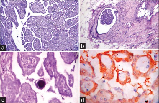
Figure 2.

(a, b, d) Photomicrograph showing histology of mesothelioma showing broad papillae with edematous fibrous cores and lined by uniform cuboidal cells with vascular invasion (b) and psammoma bodies (d), (H and E stain, ×10 view). (c) The neoplastic cells are calretinin positive (IHC stain, ×40 view)
