Figure 5.

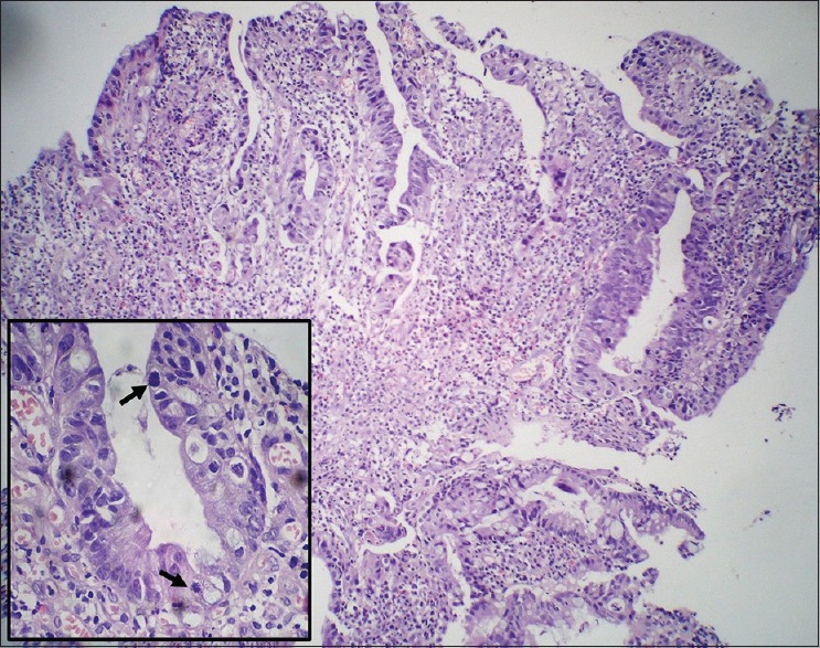
Rectal biopsy showing adenocarcinoma displaying nuclear stratification and crowding (H and E, ×40) with frequent atypical mitotic figures in inset (arrow) (×400)

Rectal biopsy showing adenocarcinoma displaying nuclear stratification and crowding (H and E, ×40) with frequent atypical mitotic figures in inset (arrow) (×400)