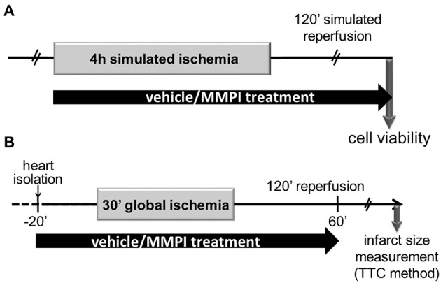
Figure 3.

Experimental protocol for cell culture studies and for the ex vivo rat heart model of AMI. (A) Isolated neonatal rat cardiac myocytes were subjected to 4 h of simulated ischemia followed by 2 h of simulated reperfusion. At the end of the reperfusion, cell viability was determined by using calcein flurescence. (B) Isolated adult rat hearts were perfused according to Langendorff and a 30-min global, no-flow ischemia was applied after a 20 min equilibration period. Subsequently, 2 h reperfusion was applied and then infarct size was determined. The hearts were perfused with Krebs-Henseleit solution containing lead candidates or vehicle from 20 min prior to the global ischemia until the 60th min of reperfusion.
