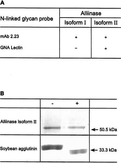
Figure 2.
A, Summary of recognition, by western blotting, of alliinase isoform I and II by monoclonal antibody 2.23 and GNA lectin. +, Recognition; −, non-recognition. B, Coomassie blue staining after SDS-PAGE of denatured alliinase isoform II and the soybean aggluinin lectin after incubation without (−) and with (+) endo H.

