
Fig. 2. Motility arrest by TRM cells after cognate antigen interaction.
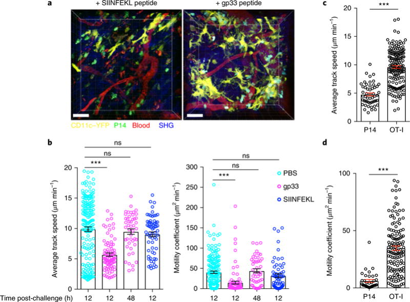
a, To assess in situ TRM cell reactivation, we transferred naive GFP+CD8+ P14 T cells into female C57BL/6J mice 1 d before infection with LCMV Armstrong, and 60 d later we challenged the mice trans-cervically with PBS, P14 reactivating peptide gp33 or control peptide SIINFEKL. Shown are representative snapshots, from Supplementary Videos 4 and 5, of the maximal projection of 3D z-stack images of P14 immune chimeras 12 h after trans-cervical exposure to peptide. Green, GFP+CD8+ P14 T cells; yellow, CD11c+ dendritic cells. Scale bars, 40 μm. b, Average track speed and motility coefficients of GFP+CD8+ P14 T cells 12 h or 48 h after trans-cervical challenge with the indicated peptide. c,d, To assess the motility of a bystander population of memory T cells, we transferred CFP+CD8+ P14 T cells into naive mice 1 d before LCMV infection. Thirty days later, we transferred GFP+CD8+ OT-I T cells into these LCMV immune chimeras, and then infected them with VSV-OVA. Thirty days after VSV-OVA infection (60 d after LCMV infection), we challenged these double-immune chimeras trans-cervically with gp33 peptide, and carried out intravital microscopy 12 h after challenge. The average track speed (c) and motility coefficients (d) of P14 and OT-I CD8+ T cells 12 h after gp33 peptide challenge are shown. Data are representative of two separate experiments with 4 mice per group per experiment. ***P< 0.001; ns, not significant; Kruskal-Wallis ANOVA with Dunn’s multiple comparison test (b) or Mann-Whitney U-test (c,d). Data in b-d are shown as the mean ± s.e.m.; circles represent individual cells.