
Fig. 6.
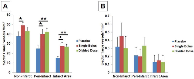
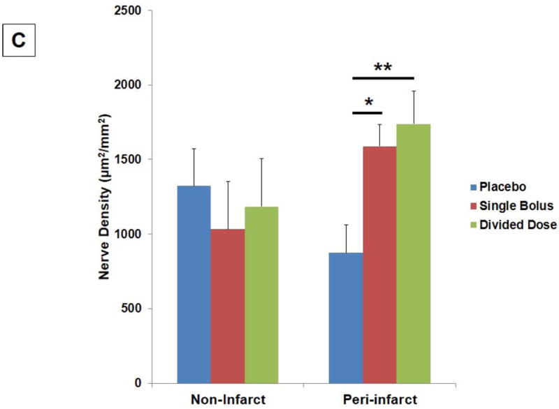
(A) Significant increases in the number of ±-actin+ small and collapsed vessels were found in the single bolus group in the non-infarct (*P<0.05), peri-infarct (*P<0.05), and infarct areas (*P<0.05). Significant increases in the number of ±-actin+ small and collapsed vessels were also found in the divided dose group in the peri-infarct (**P<0.01) and infarct areas (**P<0.01). (B) No significant differences in the number of ±-actin+ large vessels were found among 3 groups. (C) Significant increases in the GAP43+ nerve density were found in the single bolus (*P<0.05) and the divided dose group (**P=0.005) when compared to the placebo group in the peri-infarct areas.