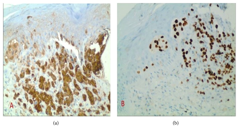
Figure 4.
Immunohistochemical stain of the scalp lesion. Positive S-100 (a) 20x, SOX-10 (b) 20x. S-100 is a cytoplasmic stain, proving the malignant cells are originating from the neural crest derived tissue (melanocytes, glial cells, and Schwann cells). SOX-10 is a nuclear stain, confirming that the malignant cells are melanocytes.

