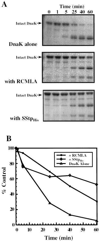
Figure 2.
Substrate protection of DnaK from trypsin degradation. A, 0.7 μm DnaK was treated with trypsin after a 5-min incubation in the presence of no unfolded protein substrate (top); 7 μm RCMLA (middle); and 7 μm SStp (bottom). Aliquots were removed at the given times and immediately boiled in SDS sample buffer. All samples were examined by SDS-PAGE and stained with Coomassie Brilliant Blue. B, Quantitation of the amount of intact DnaK remaining at each time point in A for each treatment.

