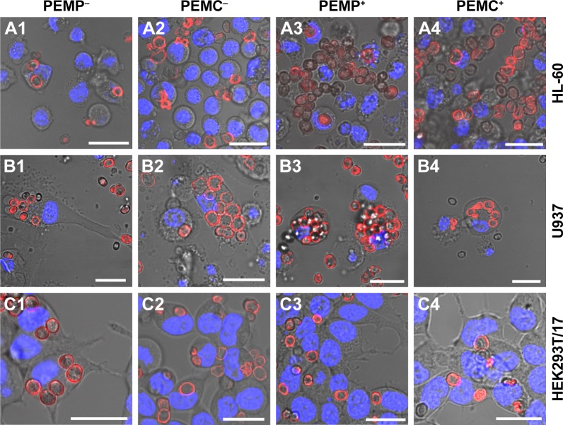
Figure 3.
Representative CLSM images after co-incubating LbL-microparticles (columns 1 and 3) and LbL-microcapsules (columns 2 and 4) with negative (columns 1 and 2) or positive (columns 3 and 4) surface charge and different cell types (A: HL-60 cells, B: U937 cells and C: HEK293T cells) to differentiate internalized LbL-microcarriers and LbL-microcarriers just adhering to the cell surface.
Note: Scale bars: 20 µm.
Abbreviations: CLSM, confocal laser scanning microscopy; LbL, layer-by-layer; PEMCs, polyelectrolyte-coated microcapsules; PEMPs, polyelectrolyte-coated microparticles.

