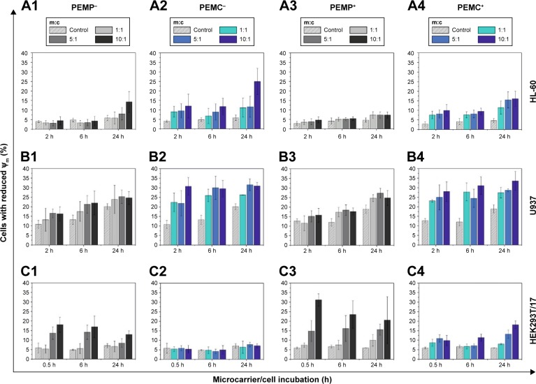
Figure 5.
Detection of early apoptotic signaling by FCM measurements after co-incubating different cell types (A: HL-60 cells, B: U937 cells and C: HEK293T cells) with LbL-microparticles (gray bars) and LbL-microcapsules (blue bars) with negative (columns 1 and 2) or positive (columns 3 and 4) surface charge in a time- and concentration-dependent way.
Notes: Cells with reduced mitochondrial membrane potential (Ψm) are referred to as apoptotic cells. All data points represent mean values with standard deviation, n≥6.
Abbreviations: FCM, flow cytometry; LbL, layer-by-layer; m:c, microcarrier:cell; PEMCs, polyelectrolyte-coated microcapsules; PEMPs, polyelectrolyte-coated microparticles.

